पत्ती
How the Incredible Evolution of Microscopes Changed Science Forever
The development of magnifying instruments spans several centuries. Optical advances gradually transformed crude glass lenses into powerful research devices. Early glass workers first crafted basic spectacles in fourteenth century Italy. Two Dutch spectacle makers created the initial compound magnification system around fifteen ninety. Robert Hooke later published his famous scientific studies in sixteen sixty five and introduced the term cells. Antonie van Leeuwenhoek then observed tiny living bacteria through single lens instruments in sixteen seventy four.
Oscar Turner
लेखक
झलक
नीचे पूरा पाठ फ़्लोट करता है।
शेल्फ़
स्टोरी लीफ़
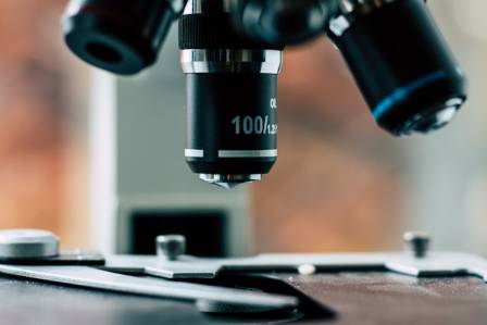
The development of magnifying instruments spans several centuries. Optical advances gradually transformed crude glass lenses into powerful research devices. Early glass workers first crafted basic spectacles in fourteenth century Italy. Two Dutch spectacle makers created the initial compound magnification system around fifteen ninety. Robert Hooke later published his famous scientific studies in sixteen sixty five and introduced the term cells. Antonie van Leeuwenhoek then observed tiny living bacteria through single lens instruments in sixteen seventy four.
Chester Moore Hall created the achromatic lens during the seventeen thirties to reduce color distortion. Joseph Jackson Lister improved compound lens arrangements in eighteen thirty to reduce spherical blurring. Ernst Abbe formulated the mathematical relationship between resolution and light wavelengths in eighteen seventy eight. Richard Zsigmondy created the ultramicroscope in nineteen hundred and three to view sub microscopic particles. Max Knoll and Ernst Ruska built the first electron microscope in nineteen thirty one. Frits Zernike invented phase contrast systems in nineteen thirty two. Gerd Binnig and Heinrich Rohrer created the scanning tunneling microscope in nineteen eighty one. Modern digital imaging systems now expand scientific research across global laboratories.
The Greek and Latin Roots of the Term
The term microscope originates from modern Latin terminology. The word combines two classic Greek terms. The prefix mikros means small. The root word skopein means to view or examine. The combined name describes an instrument designed for inspecting minute objects. Historical records show that scholars officially adopted the adjective microscopic during the seventeen sixties.
Early Visual Experiments Before True Microscopes
Early scholars experimented with magnification long before modern instruments existed. Ancient Chinese records describe water filled viewing tubes. Scholars looked through a lens at the end of a filled cylinder to inspect enlarged objects. Adjusting water levels altered the overall magnification strength. These ancient records claim scholars achieved high magnification levels several thousand years ago.
Ancient Mediterranean civilizations also documented curved lens optics. Greek scholars recorded the properties of curved glass surfaces. Young students used simple glass lenses to concentrate sunlight and spark small fires. Physicians utilized curved lenses during surgical tasks to treat medical wounds. Ancient Romans and Egyptians crafted basic curved glass pieces as well. However, those early cultures did not construct multi lens compound systems.
Optical Discoveries That Shaped Early Glass Craft
The fall of ancient Rome paused major optical progress for over a thousand years. European spectacle makers finally revived the field during the late Middle Ages. Italian artisans crafted the first wearable eyeglasses during the late thirteenth century. Italian makers argued over who created the technology first. Historical records and grave inscriptions highlight these early commercial disputes. Local monks openly praised the invention for helping aging scholars read manuscripts.
New lens grinding methods spread rapidly across Europe after the invention of the printing press. Roger Bacon analyzed optical properties extensively during the thirteenth century. Thomas Digges and Hans Lippershey expanded lens designs in England and Holland. Galileo Galilei quickly adapted these optical principles to build astronomical telescopes. Isaac Newton later constructed the reflecting telescope using curved metallic mirrors.
The Creation of the First Compound System
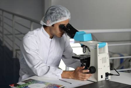
Dutch glass makers Hans Lippershey and Zacharias Janssen pioneered early compound optical systems. They aligned multiple glass lenses inside a hollow tube during the late fifteen nineties. They noticed that an image enlarged by one lens could be magnified again by a second lens.
This fundamental concept created the basic structure of the compound microscope. Early devices suffered from glass impurities and crude grinding techniques. However, the dual lens design established the groundwork for all future optical microscopy.
Robert Hooke and His Famous Scientific Illustrations
English scientist Robert Hooke popularized microscopic study during the mid seventeenth century. Hooke was a prolific inventor who developed universal joints and breathing apparatuses. He also formulated the law of elasticity in physics.
Hooke released his celebrated book Micrographia in sixteen sixty five. The book contained detailed drawings of magnified biological specimens. Readers marveled at intricate illustrations of nettle hairs, insect bodies, and thin cork slices. Hooke described the boxy cork structures as cells. That term became a foundational concept in biological science.
Antonie van Leeuwenhoek and Microscopic Organisms
Dutch draper Antonie van Leeuwenhoek made extraordinary contributions to optical science. He ground hundreds of small high quality lenses by hand. His single lens instruments achieved impressive magnification levels and remarkable image clarity.
Leeuwenhoek examined pond water samples in sixteen seventy four. He observed tiny moving organisms swimming inside the liquid drops. He described these microscopic creatures as tiny animals in detailed letters sent to scientific societies. He was the first human to witness living bacteria. Scientists took nearly two centuries to recognize cells as the basic units of life.
Lenses and Image Quality Improvements
Eighteenth century instruments suffered from blurry color distortion and physical glass imperfections. Chester Moore Hall invented the achromatic lens in the seventeen thirties to realign color wavelengths. Joseph Jackson Lister solved spherical distortion in eighteen thirty by placing weak lenses at calculated distances. Instrument makers later introduced rotating nosepieces to swap objective lenses quickly.
Optical scholar Ernst Abbe developed mathematical formulas to calculate light behavior inside microscopes. He proved that resolution depended on light wavelengths rather than simple power amplification. Abbe designed mathematical objective lenses and light condensers to focus illumination evenly onto samples. August Kohler introduced uniform lighting methods in eighteen ninety three to eliminate glare. Specialized cutting tools called microtomes allowed technicians to prepare ultra thin tissue samples. These technical improvements enabled major medical breakthroughs including pasteurization and disease identification.
The Expansion of Specialized Twentieth Century Instruments
Standard optical microscopes reached their theoretical physical resolution limit by nineteen hundred. Inventors created specialized non visible light instruments to observe smaller structures. Carl Zeiss introduced commercial ultraviolet microscopes in nineteen hundred and four to double standard resolution.
Frits Zernike invented phase contrast microscopy during the nineteen thirties. His technique rendered transparent living cells visible without chemical dyes. Max Knoll and Ernst Ruska built the transmission electron microscope in nineteen thirty one. Electron beams replaced visible light waves to achieve magnifications up to two million times. Gerd Binnig and Heinrich Rohrer created scanning tunneling microscopes in nineteen eighty one to map surfaces at atomic scales.
Diverse Modern Instrument Types
Modern microscopy features specialized tools designed for specific scientific tasks. Compound instruments inspect high magnification biological slides. Stereo units provide low magnification three dimensional views for dissections and industrial assembly. Inverted systems examine cell cultures from below the sample vessel.
Phase contrast tools illuminate transparent biological specimens without damaging live cells. Digital setups display live camera streams on electronic monitors. Modern laboratories select instruments based on specific research workflows rather than relying on one general tool.
Digital Integration and Screen Observations
Digital sensor integration transformed modern laboratory research. Modern setups replace traditional physical eyepieces with digital cameras and high resolution displays. Operators capture, record, and stream microscopic footage across computer networks.
Digital tools streamline group discussions inside university classrooms and research facilities. Technicians save high resolution files for quality control and documentation. Portable digital instruments allow field researchers to inspect outdoor samples easily.
Expanded Global Manufacturing and Availability
International manufacturing networks expanded optical accessibility dramatically in recent decades. Manufacturing facilities across Asia produce high precision components at affordable costs. Lower production expenses allow schools, hobbyists, and small businesses to purchase advanced instruments. High quality optical technology is no longer restricted to wealthy research institutions.
Why Studying Microscope Evolution Remains Valuable
Understanding the historical progress of optical science illuminates modern technical choices. Every historic advancement solved a specific physical limitation. Early pioneers corrected color distortion, increased lighting control, and pushed physical resolution limits.
Modern buyers evaluate the same core optical parameters today. Selecting an instrument requires balancing magnification power, lighting quality, resolution, and digital software needs. The long evolution of microscopic technology continues to shape modern scientific discovery.
पास की पत्तियाँ

एक बढ़ती हुई क्रांति
रियान

हेनले सिटीज रिपोर्ट के अनुसार, 2024 में दुनिया के सबसे धनी शहर
मनन

Strawberry Cream Puffs: Flaky Perfection
Nolan Drake

The Most Loved Beaches: A Trip Through America’s Waterfront Gems
Carl Allen

Faye, 5, battles snow and ice to reach the top of Ben Nevis
Freya
अपने वॉर्डरोब को परफेक्ट हैट से स्टाइल करें
ममता बस्नेत